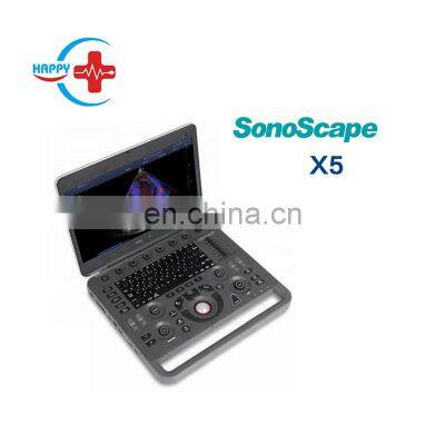
sonoscape x5 ultrasound scanner color ultrasound scan easy scanner
Product Description
Product DescriptionProduct typeB ultrasound scannerBrandSonoscapeModelX5Probe connector2Display15.6" High Resolution LED Color MonitorHard Disk500GBImaging modesB/ 2B/ 4B/ M/ CFM/ CFMM/ PDI/ DirPDI/ PW/ CW/ TDI/ AMMSonoScape’s laptop styled color Doppler ultrasound system, the X5, revolutionized the ultrasound market. Designed as the go-to ultrasound in any setting at the bedside or aboard such as in an ambulance thanks to its light weight, small size and impressive durability. Packed with crystal clear imaging capabilities and advanced applications inside this small laptop system makes it the ideal system for use inside hospitals too. Be it the OR, emergency room or ICU, the X5 can be found and ready to use at any time.Features: * Dynamic Multi-beam processing technology Dynamically provides multiple beams launching in different scanning modes, which balances parameter demands in various applications. It presents detailed information with good spatial resolution or real time movement with suitable line density and frame rate. * Spatial compound imaging X5 transmits waves from different directions, in order to get a comprehensive signal and information. It provides 9 direction lines which multiply and enhance the resolution in any depth to present sonographer more detailed information for lesion diagnosis. * Tissue Specification Imaging The system detects different tissues automatically by matching different acoustic ranges, from which the user can acquire images with more uniformity and higher spatial resolution. * C-field beam Unlike the traditional focus concentrating on limited areas, C-field beam, with a continuously dynamic focus, provides more energy contributing to better uniformity in the whole image. * SR Flow A new innovative technology, SR Flow improves the capability of detecting low velocity flow signal, while improving spatial resolution and overcoming the overflow to present user’s with real hemodynamics information.Standard Software include:Imaging modes: B/ 2B/ 4B/ M/ CFM/ CFMM/ PDI/ DirPDI/ PW/ CW/ TDI/ AMMDynamic Multi-beam TechnologyTissue Harmonic ImagingPure Inversion Harmonic ImagingTissue Specification ImagingSpatial Compound ImagingWidescan: Trapezoid ImagingConvex Extended Imagingμ-Scan: 2D speckle reduction technologyPW Auto TraceTEI IndexDICOM 3.0Auto: Auto optimization forB/ M/ PW/ CWTGC: Time gain compensationLGC: Lateral gain compensationSR FlowTriplexB/C dual liveStandby Mode2D Panoramic ImagingB Mode Prospective SavingShow Galary2D SteerVis-NeedleAuto IMTWifiStardard Configured Transducers:Linear array L741(Vascular, Small Parts, MSK etc.), 4.0-16.0MHz/ 46mmConvex array 3C-A (Abdominal, Obstetrics, Gynecology), 1.0-7.0MHz/ R50mm
>> Back Angle Steel Milling CNC Machine China Equipment
>> Powder Metallurgy Factory Supply Bronze Bushing Self-Lubricating Bearing Motor Bronze Bush
>> Fashion New Electric Bicycle 48V Comfortable Shock-Absorbing Electric Scooter Bicycle
>> Hose Crimping Machine with High-Accuracy Fi 6mm-Fi 51mm
>> High Quality Multi-purpose Bedroom Premium Foldable Bamboo Bed Laptop Desk Tray
>> Taijia electromagnetic flow meter flowmeter 12v electromagnetic flowmeter for Chemical industry
>> Premium Quality Sichuan Pepper
>> Heavy-Duty 3000lbs Permanent Magnetic Lifter for Industrial Use
>> Best Price Sale Bamboo Wood Chips Peanut Coconut Shell Burning Activated Regeneration Carbon Furnaces
>> Latest Aluminum Windows Frames Grill Design Modern Soundproof Window Solid Design
>> China manufacturer Jingxin Brand straw rope weaving machine for sale
>> 6.38 (331) mm/10.38 (551) Mmlaminated Glass/Tinted Laminated Glass/Tempered Laminated /Building Glass/Laminated Window Glass/Tempered Glass/ Shower Door Glass
>> Design Free Shipping Comfortable Leather Luxury Handbags Tote Hand Bag for Women
>> Popular Refrigerator Door Gasket Welding Machine PVC Gasket Welder Magnetic Rubber Seal Making Machine
>> PE HDPE LDPE PPR Plastic Water Gas Oil Supply Pipe Tube Extrusion Production Line Single Screw Extruder Pipe Making Machine Factory Price
>> China 5L Double Station Plastic PE Bottle Blowing Machine Factory
>> Wall-mounted humidifier
>> High Quality Wholesale Plastic Lotion Pump Cream Pump Liquid Soap Dispenser Pumps
>> Artificial Screens Table Indoor Garden Modern Glass Surround Mist Cast Iron Fireplaces
>> Kango Whole Sale Manufacturer Shoulder Cords Uniforms Silk Gold Silver Cords Security Uniforms
>> High Quality Titanium Bundle in Coils
>> Farm Machinery Tractor Mounted 3-Point Finishing Mower with Ce
>> 4000W Construction Automatic Concrete Chaser Mounted Cutting Machine for Sale
>> HDPE Flakes Plastic Pelletizing Machine for Two Stage Water Ring Cutter Plastic Recycling Granulator Line
>> Worcraft ATV Tires 22X10-10 25X8-12 25X10-12 26X9-12 26X11-12 30X10-14 32X10-14 32X10-15 33X10-15 34X10-15 32X10-16 34X10-16 34X10-18 Llanta PARA Cuatrimoto
>> Automatic Extrusion Molding Machine Plastic Toy Making Machinery
>> Chinese HDPE Poly Pex Pipe Large Diameter Plastic Product for Water Irrigation
>> Kaisen Zk-18 Crawler Truck Dumper Driven Gear Gearbox Transmission Assembly Harvester
>> High-Performance TPU Hot Melt Adhesive for Instant Bonding
>> Wholesale Sweat Suits Women's Sportswear Fitness Sports Suits For Running Sportswear Gym Track Suit Women Crop Top Set